Short project description:
Mechanical modelling of epithelial-to-mesenchymal transitions
This project is a collaboration with Eric Theveneau, Marina A. Ferreria, Diane Peurichard, Pierre Degond and Sara Merino-Aceituno.
Preprint:
- S. Plunder, C. Danesin, B. Glise, M. A. Ferreira, S. Merino-Aceituno, E. Theveneau, Modelling variability and heterogeneity of EMT scenarios highlights nuclear positioning and protrusions as main drivers of extrusion. (2023) bioRxiv.
The epithelial-to-mesenchymal transition (EMT) is a complex process during which epithelial cells[1] transition into mesenchymal cells via a progression of changes in the cell state. During this transition, fundamental cell properties change. For example, epithelial cells typical have adhesive bonds within each other. But mesenchymal cells might lose these adhesive bonds and start to migrate. However, the EMT process is very flexible in the sense that some changes might be skipped or reversed. To study this kind of heterogeneity is one of the key challenges of this project.

Image source: wikipedia.org
Project aims
The overall aim of our mathematical model is to understand the mechanical factors involved in epithelial-to-mesenchymal transition and the resulting cell migration. As there are many different directions to explore with in vivo experiments, gaining additional insight into the cell dynamics with simulations would help in the design of experiments. Ideally, the model could indicate which directions are most promising.
Concretely, we want to use simulations to come up with biological hypotheses which can be tested in vivo.
A second goal is to use the computer model also to study scenarios that are difficult or almost impossible with current experimental tools. For example, we can easily control the mixture of different cell types to model heterogeneity in computer simulations. Doing the same experimentally is only possible to a limited degree. At the same time, it is observed that cells during EMT are very heterogeneous, which raises the question if this has an actual mechanical advantage or if the heterogeneity emerges for other reasons.
The model
Without going into too many details, our model is a continuation of Marina's model where we added extra rules to model EMT.
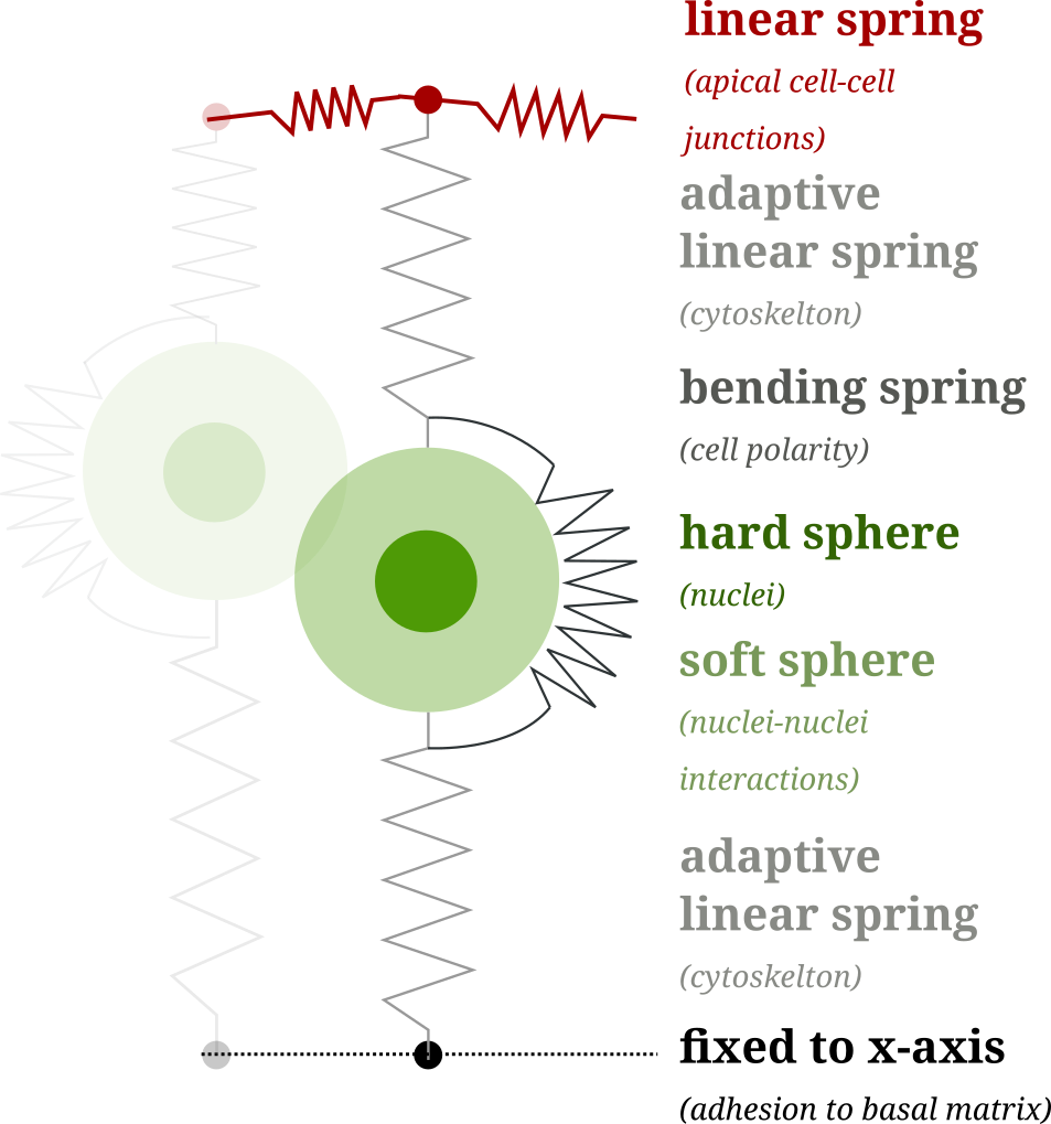
The core of the model is a "tennis ball in a sock" description of the cells: Since the cell membrane is stretched out (like a sock) and the nucleus is the hardest part of the cell (like the tennis ball) we consider the nucleus as a sphere-like object and the membrane and the other cell structure as linear springs.
Standalone EMT simulator
To see the model in action, we developed a standalone online simulator: semtor.github.io. Try it out!
Mathematical aspects
Compared to the previous model, which was a quasi-steady state simulation, we use a a first-order differential equation for the model behind simulations as the one above. Using first-order differential equations allows us to use a variety of different numerical methods. And it turned out that position-based dynamics was with quite some margin the fastest method for our model.
The model is (roughly) of the form
The degrees of freedom are the positions of the nuclei and the upper and lower endpoints of the cell membrane (which we call apical and basal points). The mathematically inclined reader might notice that the inequality constraints add some extra challenges. But we will not go into these details here.
In addition to the differential equation, there are many additional rules which change certain properties of the model at discrete times. For example:
- Cell division,
- Growth of the cell before division,
- Typical for pseudo-stratified tissue: The cytoskeleton pulls the nucleus towards the apical layer, just before the division starts.
The EMT model
To model EMT we added the following events
- A: Loss of apical adhesion (removal of apical-nuclei spring),
- B: Loss of basal adhesion (removal of basal-nuclei spring),
- S: Degeneration of cell straightness (reduction of the bending spring),
- p: Protrusions (cells start moving in direction of their polarity).
In a simplified world, cells would perform
Results
A full display of all aspects of the model is beyond the scope of this brief project description.[2] To show a one aspect, I will present one simulation of cells that lose their apical adhesion (after 12h) and are then passively pushed towards the basal side.
In the video, the white cells are from the control population without EMT and the red cells perform EMT.
Some observations which can be made in the model:
- The loss of adhesion removes the EMT cells from the center of the tissue.
- After EMT, most of the cells are positioned on the basal side of the tissue (black line), from where they could further migrate through the basal matrix.
- Some EMT cells extrude towards the apical side (red line) of the tissue. This can be avoided by adding protrusive activity during EMT.
Numerical aspects
The numerical simulation of differential equations with inequality constraints is an old but very diverse topic. The preferred numerical methods vary fundamentally depending on the application, ranging from discrete element methods for engineering applications to complex algorithms from non-smooth mechanics or elasticity.
Our method of choice is the rather novel position-based dynamics (PBD). The advantage of PBD is that it has nearly optimal speed and it does not waste unnecessary resources on satisfying the inequality constraints. This allowed us to run millions of simulations within a few hours. With this uncompromising approach, we could generate enough data for statistical data analysis. Moreover, the numerical stability of PBD resolved issues previous algorithms had when the tissue is very packed.
To be continued...
This description is about ongoing research, as such, it is subject to change and none of the results is final.